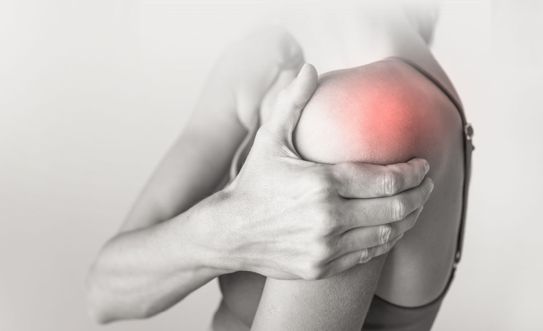
Shoulder Surgery At Acumen.
Dr. Jesse Slade Shantz- Private surgery in Calgary and Vancouver.
Dr. Curtis Myden- Private surgery in Edmonton.
For any Canadian, in or out of Province.
Shoulder Diagnosis
The main pain generators in the shoulder are usually the rotator cuff, long head of biceps tendon, the acromioclavicular joint and the glenohumeral joint including the labrum and joint capsule. A detailed exam with an experienced shoulder specialist is the first step to determining the cause of shoulder problems.
Imaging of the Shoulder
There are several ways to investigate shoulder issues using imaging, and each shoulder problem demands a different approach.
X-ray
These can be of the shoulder, humerus or can focus on the acromioclavicular joint (AC joint).
Ultrasound
The soft tissue of the shoulder can be seen using ultrasound imaging. The rotator cuff, biceps tendon and AC joint can all be seen using this technology.
Ultrasound is also used at Acumen Health for any injections, ensuring accuracy.
CT Scan
These cross-sectional scans provide detailed information about the bony structure of the shoulder. The images can be used to create 3D models of the shoulder joint to help with surgical planning in cases of glenohumeral arthritis and instability where bone loss is a concern.
Standard MRI
For the imaging of the rotator cuff a standard MRI provides significant details. MRI is particularly useful in the assessment of rotator cuff muscles in cases of rotator cuff tears where there is muscle wasting.
MRI Arthrogram (MRA)
In this more invasive test gadolinium dye is injected into the glenohumeral joint by a doctor before an MRI. This highlights the capsule of the shoulder joint and help to better visualize the labrum. These exams are most useful in cases of instability or SLAP tears of the long head of the biceps.
Shoulder Surgery
Shoulder surgery addresses four main issues that can affect shoulder function: pain, weakness, instability/dislocations and stiffness or lack of movement. The majority of shoulder surgery can be accomplished arthroscopically, which involves using small incisions to place a camera and specially designed tools into the shoulder joint in order to correct problems.
For some diagnoses, open shoulder surgery is required such as some cases of long head of biceps, pectoralis major tears, shoulder instability with bone loss or failure of previous arthroscopic stabilization and shoulder replacements for arthritis.
Many shoulder surgeries are performed using nerve blocks to limit pain. In addition, the majority of patients also have general anaesthesia during the procedure. .
SHOULDER SURGICAL OPTIONS
Rotator Cuff Repair | Labral Repairs | Shoulder Stabilization | Shoulder Replacement | Shoulder Arthroscopy | Rotator Cuff Tears | Shoulder Impingement | Shoulder Dislocations | Multidirectional Shoulder Instability Shoulder Tendinitis | Conditions | Shoulder | Osteoarthritis Biceps Pathology | Frozen Shoulder | SLAP Lesions Labrum Tears | Joint
Common Shoulder Injuries
Book an appointment with us
This form should not be used to enter any health information. The security of any health information cannot be guaranteed.


